Perhatikan gambar proses megasporogenesis berikut!...
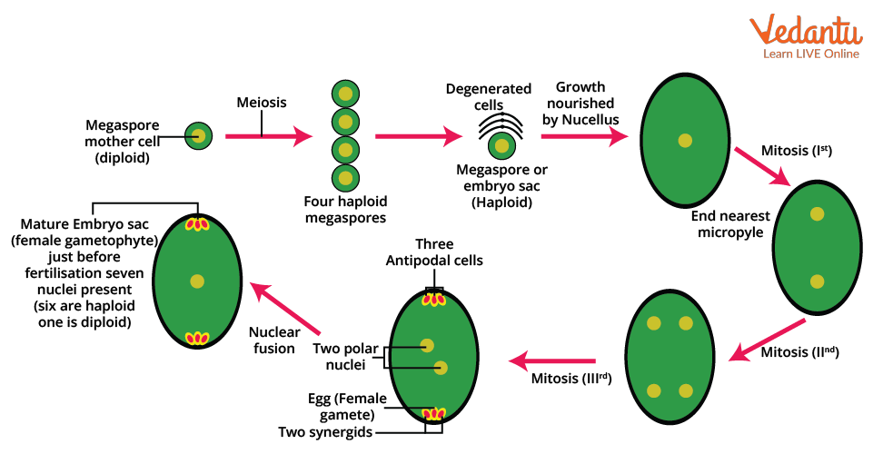
Megasporogenesis starts with the specification of the megaspore mother cell (MMC). After MMC meiosis produces four haploid megaspores, one functional megaspore persists, while the remaining megaspores degenerate (Figure 1) [ 38 ]. Little is known about the molecular mechanisms underpinning monospory and functional megaspore selection; although.

Megasporogenesis & Embryo sac development, Monosporic & Bisporic types YouTube
In this work megasporogenesis, megagametogenesis and aril ontogeny were studied in Cytisus multiflorus and C. striatus, endemics from the western Mediterranean region. • Methods Ovaries and ovules from flower buds, flowers at anthesis and hand cross-pollinated flowers were sectioned with a rotary microtome and studied under light and.
Megasporogenesis Introduction, Structure, Process and FAQs
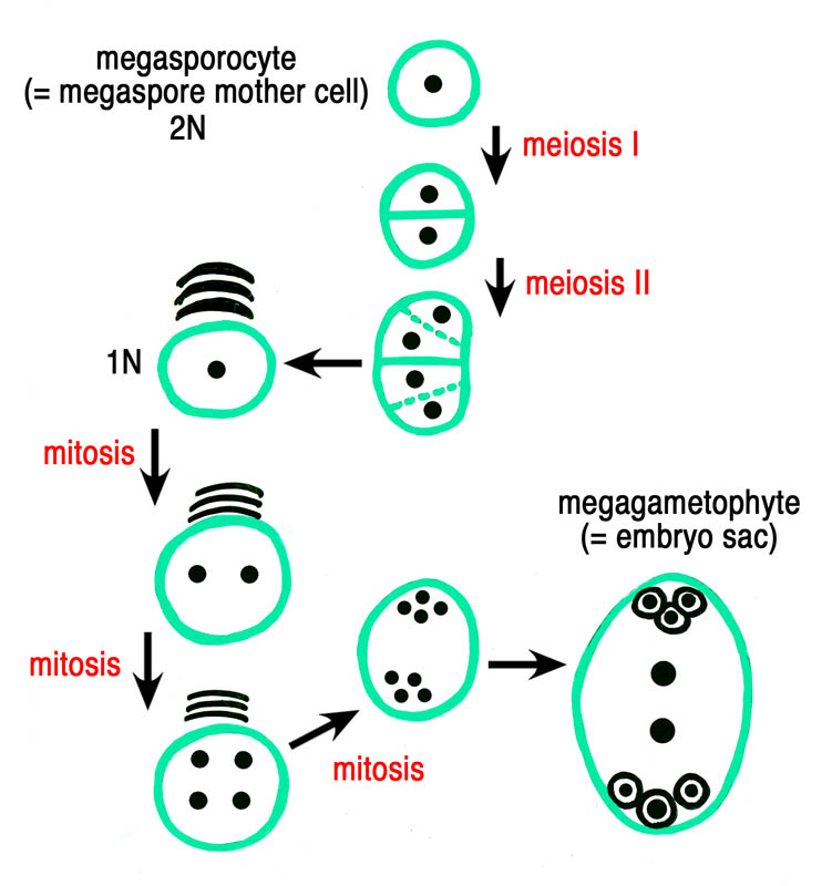
Megasporogenesis refers to the development of megaspores from the megasporocyte, the cell that undergoes meiosis. Meiosis of the megasporocyte nucleus results in the formation of four haploid megaspore nuclei. In most taxa, meiosis is followed by cytokinesis, resulting in four megaspore cells. This pattern is termed monosporic megasporogenesis.

ボード「Fruit & seeds」のピン
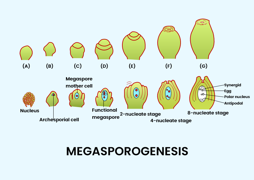
Megasporogenesis is the term used to describe the process that results in the formation of megaspores. Megaspore mother cells undergo a process called meiosis or reduction division to produce these megaspores. A single cell within the nucellus, located at the micropylar region, differentiates into a megaspore mother cell.

MEGASPOROGENESIS [CLASSXII BIOLOGY]ZOOM RECORDED CLASS YouTube
The carpel is the gross morphological part of the flower concerned with female sporogenesis and gametogenesis and is the homologue of the stamen. In its simplest form, the carpel is a leaflike organ composed of the ovary, style, and stigma, although various kinds of fusions between carpels have produced a complex organ in the modern flower.

FG formation. A Schematic presentation of megasporogenesis and... Download Scientific Diagram
Learn for free about math, art, computer programming, economics, physics, chemistry, biology, medicine, finance, history, and more. Khan Academy is a nonprofit with the mission of providing a free, world-class education for anyone, anywhere.
7 Celled 8 Nucleate Structure Ovule With Labelled Diagram
Types of Embryo Sac Development. 1. Monosporic. In the majority of angiosperms, the embryo sac develops from one functional megaspore while the other three degenerate. For example, Oenothera, Polygonum. 2. Bisporic. In this type, two of the megaspores take part in embryo sac development. Examples are Allium, Scilla, and Trillium.
Biology THE PISTIL, OVULE AND EMBRYO SAC
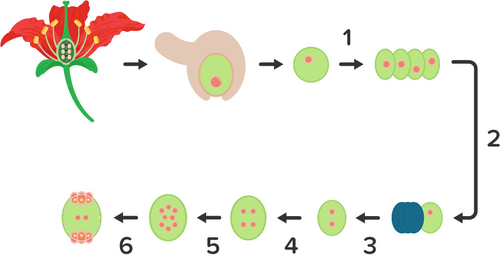
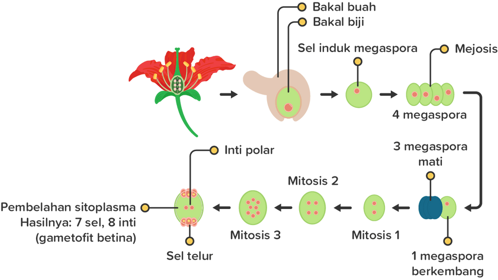
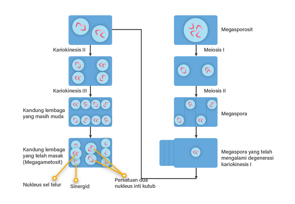
Gambar di bawah ini adalah proses megasporogenesis dan megagametogenesis dalam satu siklus. Setelah 8 inti sel tersebut menempati tempatnya masing-masing, akan mulai terbentuk dinding sel yang memisahkannya dengan inti sel yang lain. Dinding sel ini akan memisahkan masing-masing inti sel kecuali pada inti kandung lembaga sekunder yang tetap.

Megasporogenesis & Megagametogenesis Biology Class 12 NEET
Abstract. Megasporogenesis and megagametogenesis lead to the formation of the embryo sac. It includes the development of a highly differentiated plantlet in function of the sexual reproduction of angiosperms. After a meiotic division and cell isolation the development from the unicellular stage to the multicellular embryo sac represents the.
Perhatikan gambar proses megasporogenesis berikut!...
Megagametogenesis is the process of maturation of the female gametophyte, or megagametophyte, in plants. [1] During the process of megagametogenesis, the megaspore, which arises from megasporogenesis, develops into the embryo sac, which is where the female gamete is housed. [2] These megaspores then develop into the haploid female gametophytes. [2]

FG formation. A Schematic presentation of megasporogenesis and... Download Scientific Diagram
Megasporogenesis Diagram. Above is the detailed megasporogenesis flow chart, and the following are the steps: The process of creating haploid megaspores from a diploid megaspore mother cell is known as megasporogenesis (MMC). A large diploid (2n) cell known as the megaspore mother cell (MMC) conducts meiotic division to produce four haploid.
gambarlah megasporogenesis pada tumbuhan...
The essence of megasporogenesis is the formation of megaspores by meiosis of the megasporocyte (the megaspore mother cell). The transformation of the megaspore into the female gametophyte (the embryo sac housing the egg) is the centerpiece of the subsequent events of female sexual differentiation in flowering plants.
In angiosperms, functional megaspore develops into(a) Embryo sac(b) Ovule(c) Endosperm (d
The ovule ontogenesis and the megasporogenesis events were studied under bright field, fluorescence and scanning electron microscopy. The primordium is 3-zonate and gives rise to a hemianatropous.

MEGASPOROGENESIS & MEGAGAMETOGENESIS
Megasporogenesis, megagametogenesis and embryogenesis of Liparis elliptica (family Orchidaceae, tribe Malaxideae, subtribe Malaxidinae) have been studied. It was shown that the L. elliptica embryo sac is monosporic and develops from the chalazal cell of the megaspore triad according to the modified Polygonum type. The embryo sacs are reduced to four-six nuclei. The suspensor is unicellular.

Megasporogenesis and embryo sac development in Dioscorea caucasica. Download Scientific Diagram
Ukuran Gamet. Mikrospora hasil mikrosporogenesis berukuran kecil, namun megaspora hasil megasporogenesis memiliki ukuran yang besar. Pada bunga terlihat serbus sari berbentuk kecil dan banyak, namun bakal biji hanya ada satu dengan ukuran yang besar. Halaman Berikutnya.

I Biology Pembelahan Sel
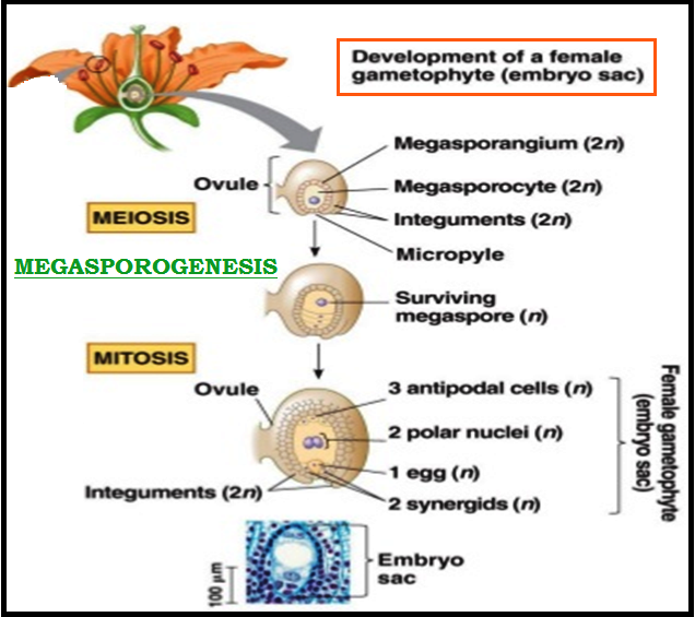
Megasporogenesis is defined as the formation of haploid megaspores by the meiotic division of diploid megaspore mother cells (MMC) inside the megasporangium (ovule). The haploid megaspore undergoes repeated mitotic divisions to form the embryo sac. During fertilisation, the haploid male gamete (present inside the pollen grain) and the haploid.